 Disease Modifying Anti-Rheumatic Drugs (DMARDs) are the only drugs demonstrated to modify the course of the rheumatoid arthritis and improve radiologic findings. There are 2 types of DMARDs: biologic and syntethic small molecules. Among the biologic DMARDs are tumor necrosis factor alpha (TNF-a) inhibitors; T-cell costimulatory blocking agents and B-cell depleting agents
Novel Treatments for Rheumatoid Arthritis
 Disease Modifying Anti-Rheumatic Drugs (DMARDs) are the only drugs demonstrated to modify the course of the rheumatoid arthritis and improve radiologic findings. There are 2 types of DMARDs: biologic and syntethic small molecules. Among the biologic DMARDs are tumor necrosis factor alpha (TNF-a) inhibitors; T-cell costimulatory blocking agents and B-cell depleting agents
Empyema With Organized Granulation Tissue
What is the definition of empyema? Why might the term organized granulation tissue be redundant? Why might empyema and granulation be oxymoronic?
Demonstrate fibrin. Demonstrate neutrophils. Demonstrate abundant new ingrowth of blood vessels.
Thoracoscopy for stage 3 organised empyema / Decortication, Delhi, NOIDA,NCR India
Thoracoscopic Decortication for stage3 organized empyema. Video showing
the procedure with removal of thick fibrous pleural layer trapping the
lung.
Thoracoscopic Surgery (decortication) for pulmonary kochs & Pyothorax
Thoracoscopic Surgery (decortication) for pulmonary kochs & Pyothorax is a video assisted laparoscopic surgical procedure which was performed by Dr. Mohit Bhandari, Director, Bhandari Hospital & Research Centre & assisted by Dr. Milind Joshi. Dr. Mohit Bhandari is also the Director of Sri Aurobindo Medical College & LASER (Laparoscopy Academy of Surgical Education & Research), Indore.
Thoracoscopy VATS Decortication in stage 2 empyema, Delhi, Noida, India.
Thoracoscopy Decortication for stage 2 / fibrinopurulent satge empyema
in a patient with parapneumonic effusion on tracheostomy for
longstanding ventilatory support with failure to decanulate
tracheostomy. CT thorax showed left multiloculated collection with lower
lobe collapsed and non draining chest tube. Patient underwent
decortication of the entrapped lung and drainage of multiloculated
fluid.
Post decortication the lung was fully expanded and the tracheostomy tube was decanulated on 3rd day.
Post decortication the lung was fully expanded and the tracheostomy tube was decanulated on 3rd day.
Histology of Thyroid
 Thyroid gland consists of colloid follicles which are lined by cuboid epithelial cells secreting the thyroid hormones, triiodothyronine (T3) and thyroxine (T4). Between the follicles is the interstitial space, containing the blood vessels and the parafolliclar (C) cells responsible for the secretion of calcitonin.
Hematoxylin and Eosin stain
 HE stain is a popular staining method in histology. Its a combination of two dyes the basic dye (hematoxylin) and the
alcohol-based dye (eosin). In an HE stain you will usually see both
eosinophilia and basophilia the nuclei of cells basophilic (blue),
while eosinophilia is typical of cytoplasmic constituents (pink).
Xylene, alcohols, distilled water are also required.
HE stain is a popular staining method in histology. Its a combination of two dyes the basic dye (hematoxylin) and the
alcohol-based dye (eosin). In an HE stain you will usually see both
eosinophilia and basophilia the nuclei of cells basophilic (blue),
while eosinophilia is typical of cytoplasmic constituents (pink).
Xylene, alcohols, distilled water are also required.MHC genes and molecules
The imune system MHC class I and II molecules and the genes that encode their synthesis are showed in this video, including an illustration of the MHC gene locus and an illustration of the MHC molecular structure.The distribution of the MHC molecules on the cells is also illustrated.
NEJM - Repositioning Dislocated Temporomandibular Joints
The temporomandibular joints are anterior to the ear. They are
ginglymoarthrodial joints that permit both rotational and sliding
movements. The temporomandibular joints, which are on each side of the
lower jaw, join the mandible to the viscerocranium and allow the mouth
to open and close.
Shoulder Anatomy Animated Tutorial
In this episode of eOrthopodTV, orthopaedic surgeon Randale C. Sechrest,
MD narrates an animated tutorial on the basic anatomy of the shoulder.
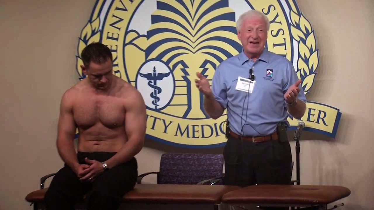
Shoulder Exam - Dr. Hawkins
Basic Wound Care - Clinical Skills
This video - produced by students at Oxford University Medical School in
conjunction with the faculty - demonstrates the principles and
techniques underlying basic wound care. It is part of a series of videos
covering clinical skills.
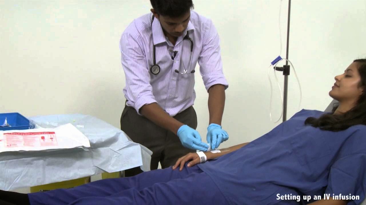
Blood Transfusion And Intravenous Infusion - Clinical Skills
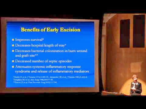
Burn Treatment - How to Achieve an LD50 of 98% TBSA Burn
Tina L. Palmieri, MD, FACS, FCCM Associate Professor and Director at
University of California Davis and Assistant Chief of Burns at Shriners
Hospital for Children Northern California, presenting lecture "How to
Achieve an LD50 of 98% TBSA Burn: Are We There Yet?", in August 3, 2011
at The 9th Annual John F. Hansbrough Memorial Lecture, Goldberg
Auditorium, UCSD Moores Cancer Center, La Jolla, California
Draining Bulla On Palm
Thoracoscopic (VATS) wedge resection of Fungal Ball (Mucormycosis) Left Upper Lobe
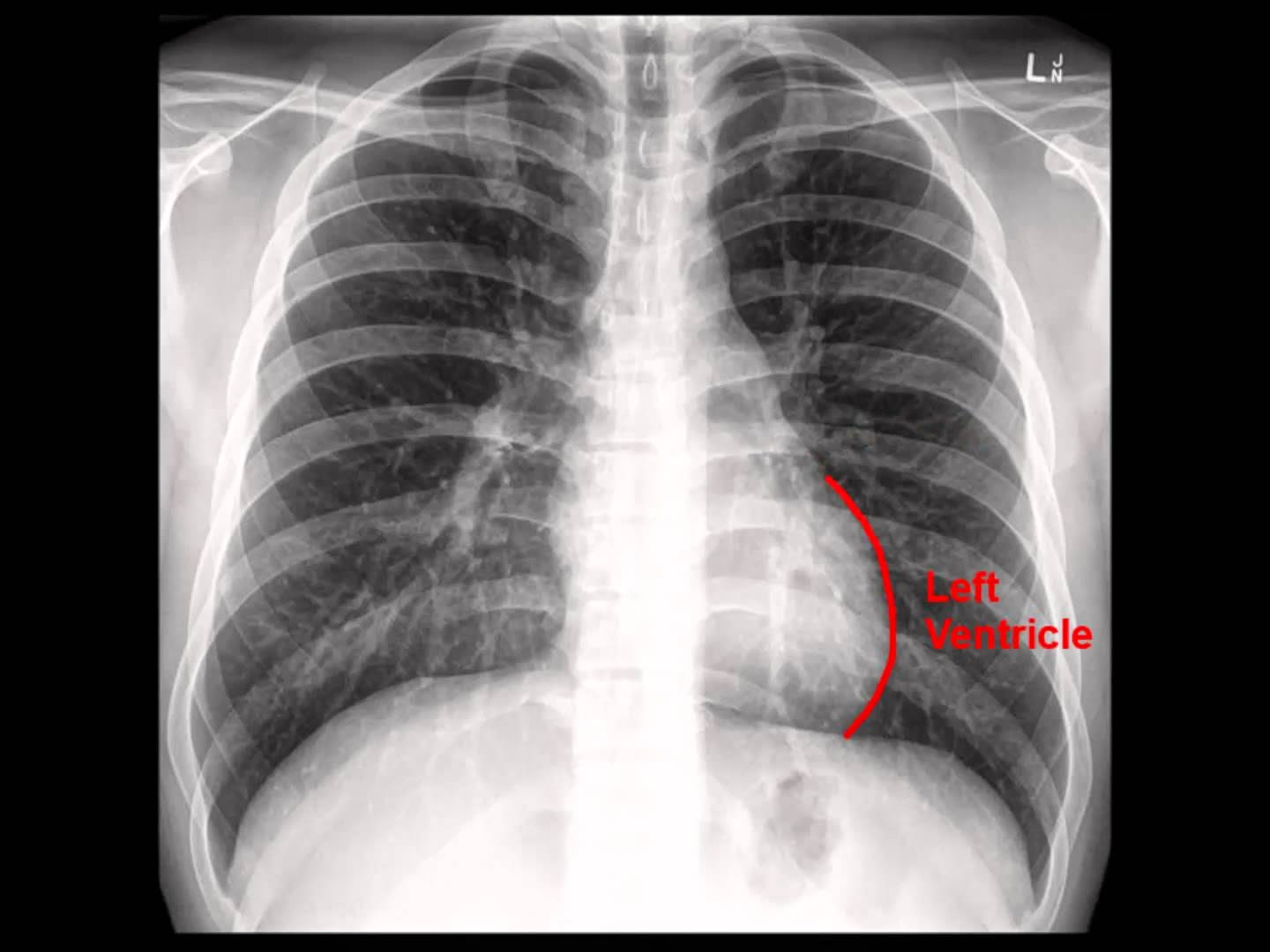
Chest X-ray: Analysis in a nutshell
Video illustrating a systematic approach to the interpretation of chest radiography. Exemplifies the most important steps such as checking the patients information, identifying the type of film used and the technical quality of the film. Also exemplifies the alphabet system, which is the most commonly used and requires analysis of the: Airways, Bones, Cardiac silhouette, Diaphragm, Edge of the heart, Fields of the lung, Gastric bubble, Hila and Instruments (A,B,C,D,E,F,G,H,I).
Layers of the epidermis
 Lecture showing the 5 layers of the epidermis: basale, spinosum, granulosum, lucidum, corneum. The layers are showed on diagrams and in pictures taken by the use of light microscopy. The cells of the epidermis are also showed: Keratinocytes, Melanocytes, Merkell cells, Langherhans cells. Presents the difference between thick skin and thin skin.
 Laparoscopic Lysis of Abdominal Adhesions
This 3D medical animation shows how adhesions in the abdomen may cause complications. These problems may include obstruction, twisting, and dislocating areas of the small intestine. Adhesions can be separated with laparoscopic instruments.
Tuberculosis and Hypervascularity video - Animation by Cal Shipley, M.D.
Chest x-ray , pneumonia
Video will describe how pneumonia may look like on a chest x-ray. Subtle
pneumonia. Please see disclaimer on my website.
www.academyofprofessionals.com. Multiple choice questions are also
availabe for those who might be interested in enhancing and testing
their knowlege.
Inguinal hernia
This 3D medical animation depicts the surgical repair of a direct,
strangulated inguinal hernia. The animation of the surgery shows the
placement of the laparascopic devices, location of the herniated
abdominal mass, and placement of mesh graft to strengthen the fascia of
the abdominal wall. ANCE00200
Surgical debridement
This 3D animation provides an overview of how dead tissue can be
removed from around a wound. Wounds can result from a variety of causes
including: injuries, burns, bedsores, decubitus ulcers and infected
surgical incisions. Dead tissue can delay wound healing and lead to
infections.
Burns: Classification and Treatment
This 3D medical animation shows burns, which are skin injuries caused by heat, chemicals, electricity, or radiation. Types of burns are shown, including first-degree/superficial burns, second-degree/partial thickness burns, third-degree/full thickness burns, and fourth-degree burns. Treatments for burns are described, including antibiotic ointments, bandages, and medications such as acetaminophen and ibuprofen.
A Medical Career - Why Not Pathology?
Beyond the Data: Preventing Adverse Health Effects from Nanotechnology
In this CDC Grand Rounds - Beyond the Data video, Dr. John Iskander and
Dr. Paul Schulte discuss the emergence of nanotechnology from science
fiction to real life: How small is small when talking about
nanotechnology, concerns for workers who handle nanomaterial, and
prevention strategies for combating health concerns.
Laparoscopic Repair of Para Umbilical Hernia performed Dr R K Mishra
This video demonstrate Laparoscopic Repair of Para Umbilical Hernia
performed by Dr. R.K. Mishra at World Laparoscopy Hospital. Ventral
hernia results from a weakness in the musculoaponeurotic layer of the
anterior abdominal wall. Many this type of hernia has root of
development during the period of development like; omphalocele,
gstroschisis and congenital umbilical hernia.
Subscribe to:
Comments (Atom)


+for+pulmonary+kochs+&+Pyothorax.jpg)




+wedge+resection+of+Fungal+Ball+(Mucormycosis)+Left+Upper+Lobe.jpg)
