New templates on your Email

Showing posts with label lung. Show all posts

Pleural biopsy in tuberculous pleural effusion

Pleural biopsy performed in a 35 year old Fijian female presenting with bilateral pleural effusion. Both classic hematoxylin & eosin stain and acid-fast staining have been performed and are illustrated in this presentation 

Empyema With Organized Granulation Tissue

http://pathologyvideos.blogspot.ro/2014/04/empyema-with-organized-granulation.html
What is the definition of empyema? Why might the term organized granulation tissue be redundant? Why might empyema and granulation be oxymoronic? Demonstrate fibrin. Demonstrate neutrophils. Demonstrate abundant new ingrowth of blood vessels.

Thoracoscopy for stage 3 organised empyema / Decortication, Delhi, NOIDA,NCR India

http://pathologyvideos.blogspot.ro/2014/04/thoracoscopy-for-stage-3-organised.html
Thoracoscopic Decortication for stage3 organized empyema. Video showing the procedure with removal of thick fibrous pleural layer trapping the lung.

Thoracoscopic Surgery (decortication) for pulmonary kochs & Pyothorax

http://pathologyvideos.blogspot.ro/2014/04/thoracoscopic-surgery-decortication-for.html
Thoracoscopic Surgery (decortication) for pulmonary kochs & Pyothorax is a video assisted laparoscopic surgical procedure which was performed by Dr. Mohit Bhandari, Director, Bhandari Hospital & Research Centre & assisted by Dr. Milind Joshi. Dr. Mohit Bhandari is also the Director of Sri Aurobindo Medical College & LASER (Laparoscopy Academy of Surgical Education & Research), Indore.

Thoracoscopy VATS Decortication in stage 2 empyema, Delhi, Noida, India.

http://pathologyvideos.blogspot.ro/2014/04/thoracoscopy-vats-decortication-in.html
Thoracoscopy Decortication for stage 2 / fibrinopurulent satge empyema in a patient with parapneumonic effusion on tracheostomy for longstanding ventilatory support with failure to decanulate tracheostomy. CT thorax showed left multiloculated collection with lower lobe collapsed and non draining chest tube. Patient underwent decortication of the entrapped lung and drainage of multiloculated fluid.
Post decortication the lung was fully expanded and the tracheostomy tube was decanulated on 3rd day.

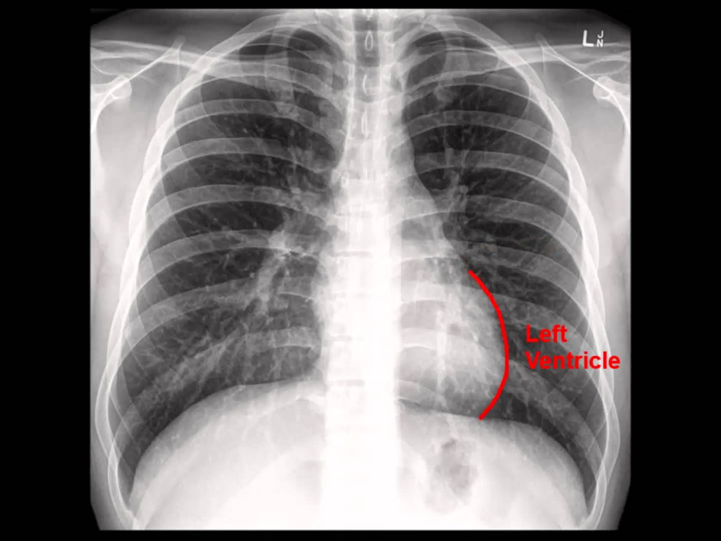
Chest X-ray: Analysis in a nutshell

http://pathologyvideos.blogspot.ro/2014/04/chest-x-ray-analysis-in-nutshell.html
Video illustrating  a systematic approach to the interpretation of chest radiography. Exemplifies the most important steps such as  checking the patients information, identifying the type of film used and the technical quality of the film. Also exemplifies the alphabet system, which is the most commonly used and requires analysis of the: Airways, Bones, Cardiac silhouette, Diaphragm, Edge of the heart, Fields of the lung, Gastric bubble, Hila and Instruments (A,B,C,D,E,F,G,H,I).

Chest x-ray - Idiopathic Pulmonary Fibrosis

http://pathologyvideos.blogspot.ro/2014/04/chest-x-ray-idiopathic-pulmonary.html A brief video on IPF (IdiopathicPulmonary Fibrosis). X-ray with IPF is courtesy of Dr Alex Maclennan and Normal Chest x-ray is courtesy of a youtube viewer.

Chest x-ray , pneumonia

http://pathologyvideos.blogspot.ro/2014/04/chest-x-ray-pneumonia.html
Video will describe how pneumonia may look like on a chest x-ray. Subtle pneumonia. Please see disclaimer on my website. www.academyofprofessionals.com. Multiple choice questions are also availabe for those who might be interested in enhancing and testing their knowlege.