 Disease Modifying Anti-Rheumatic Drugs (DMARDs) are the only drugs demonstrated to modify the course of the rheumatoid arthritis and improve radiologic findings. There are 2 types of DMARDs: biologic and syntethic small molecules. Among the biologic DMARDs are tumor necrosis factor alpha (TNF-a) inhibitors; T-cell costimulatory blocking agents and B-cell depleting agents
Novel Treatments for Rheumatoid Arthritis
Disease Modifying Anti-Rheumatic Drugs (DMARDs) are the only drugs demonstrated to modify the course of the rheumatoid arthritis and improve radiologic findings. There are 2 types of DMARDs: biologic and syntethic small molecules. Among the biologic DMARDs are tumor necrosis factor alpha (TNF-a) inhibitors; T-cell costimulatory blocking agents and B-cell depleting agentsPleural biopsy in tuberculous pleural effusion

Empyema With Organized Granulation Tissue

Thoracoscopy for stage 3 organised empyema / Decortication, Delhi, NOIDA,NCR India

Thoracoscopic Surgery (decortication) for pulmonary kochs & Pyothorax
+for+pulmonary+kochs+&+Pyothorax.jpg)
Thoracoscopy VATS Decortication in stage 2 empyema, Delhi, Noida, India.

Post decortication the lung was fully expanded and the tracheostomy tube was decanulated on 3rd day.

Histology of Thyroid
 Thyroid gland consists of colloid follicles which are lined by cuboid epithelial cells secreting the thyroid hormones, triiodothyronine (T3) and thyroxine (T4). Between the follicles is the interstitial space, containing the blood vessels and the parafolliclar (C) cells responsible for the secretion of calcitonin.
Hematoxylin and Eosin stain
 HE stain is a popular staining method in histology. Its a combination of two dyes the basic dye (hematoxylin) and the
alcohol-based dye (eosin). In an HE stain you will usually see both
eosinophilia and basophilia the nuclei of cells basophilic (blue),
while eosinophilia is typical of cytoplasmic constituents (pink).
Xylene, alcohols, distilled water are also required.
HE stain is a popular staining method in histology. Its a combination of two dyes the basic dye (hematoxylin) and the
alcohol-based dye (eosin). In an HE stain you will usually see both
eosinophilia and basophilia the nuclei of cells basophilic (blue),
while eosinophilia is typical of cytoplasmic constituents (pink).
Xylene, alcohols, distilled water are also required.MHC genes and molecules

NEJM - Repositioning Dislocated Temporomandibular Joints

Shoulder Anatomy Animated Tutorial

Shoulder Exam - Dr. Hawkins
 Dr. Richard Hawkins with the Steadman Hawkins Clinic of the Carolinas
demonstrates a comprehensive shoulder exam. This demonstration was done
during the 8th annual Sports Medicine Symposium on June 10th 2011.
©Hawkins Foundation 2011
Dr. Richard Hawkins with the Steadman Hawkins Clinic of the Carolinas
demonstrates a comprehensive shoulder exam. This demonstration was done
during the 8th annual Sports Medicine Symposium on June 10th 2011.
©Hawkins Foundation 2011Basic Wound Care - Clinical Skills

Blood Transfusion And Intravenous Infusion - Clinical Skills
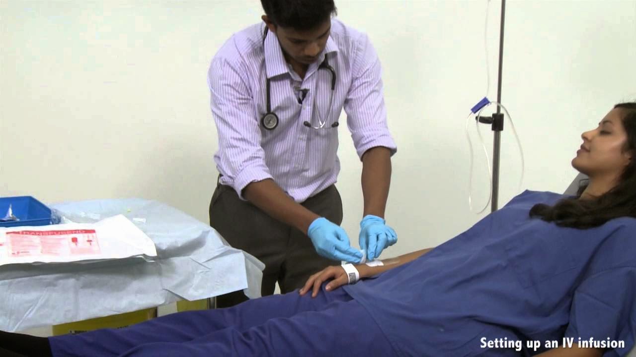 This video - produced by students at Oxford University Medical School in
conjunction with the faculty - demonstrates the principles and
techniques underlying intravenous infusion of fluids and the safe
transfusion of blood. It is part of a series of videos covering clinical
skills.
This video - produced by students at Oxford University Medical School in
conjunction with the faculty - demonstrates the principles and
techniques underlying intravenous infusion of fluids and the safe
transfusion of blood. It is part of a series of videos covering clinical
skills.Intramuscular And Subcutaneous Injections - Clinical Skills
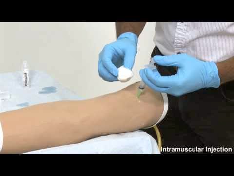 This video - produced by students at Oxford University Medical School in
conjunction with the faculty - demonstrates the principles and
techniques underlying intramuscular and subcutaneous injections.It is
part of a series of videos covering clinical skills
This video - produced by students at Oxford University Medical School in
conjunction with the faculty - demonstrates the principles and
techniques underlying intramuscular and subcutaneous injections.It is
part of a series of videos covering clinical skills Burn Treatment - How to Achieve an LD50 of 98% TBSA Burn
Draining Bulla On Palm
 Here you will see drainage of bulla on right palm. You will see the
purulent material coming out when incision was given. Watch out whole
procedure of giving incision, cleaning wound, with normal saline,
hydrogen peroxide, betadiene, & removing dead skin.
Here you will see drainage of bulla on right palm. You will see the
purulent material coming out when incision was given. Watch out whole
procedure of giving incision, cleaning wound, with normal saline,
hydrogen peroxide, betadiene, & removing dead skin.Thoracoscopic (VATS) wedge resection of Fungal Ball (Mucormycosis) Left Upper Lobe
+wedge+resection+of+Fungal+Ball+(Mucormycosis)+Left+Upper+Lobe.jpg) This is an educational video demonstrating Thoracoscopic (VATS) wedge
resection of Fungal Ball (Mucormycosis) Left Upper Lobe by Prof. Arvind
Kumar and Dr. Belal Bin Asaf, Institute of Robotic Surgery at Sir Ganga
Ram Hospital, New Delhi, India.
This is an educational video demonstrating Thoracoscopic (VATS) wedge
resection of Fungal Ball (Mucormycosis) Left Upper Lobe by Prof. Arvind
Kumar and Dr. Belal Bin Asaf, Institute of Robotic Surgery at Sir Ganga
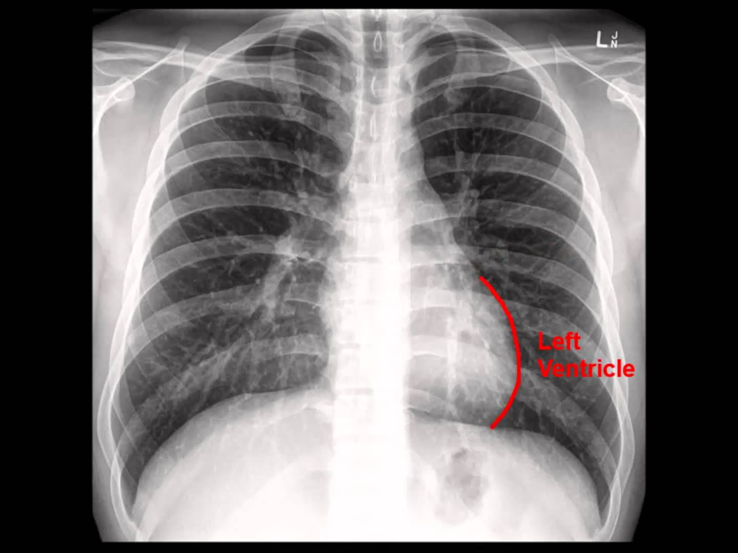
Ram Hospital, New Delhi, India. Chest X-ray: Analysis in a nutshell
Layers of the epidermis
 Lecture showing the 5 layers of the epidermis: basale, spinosum, granulosum, lucidum, corneum. The layers are showed on diagrams and in pictures taken by the use of light microscopy. The cells of the epidermis are also showed: Keratinocytes, Melanocytes, Merkell cells, Langherhans cells. Presents the difference between thick skin and thin skin.
 Laparoscopic Lysis of Abdominal Adhesions

Basic Medical Pathology: Morphological Expressions of Cell Injury
 The video tape defines and describes injuries sustained by cells when
they are exposed to stress and are unable to adapt. The results of cell
injury are shown with photomicrographs, organ photographs, and electron
micrographs of normal and injured cells.
The video tape defines and describes injuries sustained by cells when
they are exposed to stress and are unable to adapt. The results of cell
injury are shown with photomicrographs, organ photographs, and electron
micrographs of normal and injured cells.Creating skin grafts for burn victims
 Biomedical engineers at Stevens are making more realistic skin grafts by adding hairs to artificial skin tissues.
Tuberculosis and Hypervascularity video - Animation by Cal Shipley, M.D.
 Infection of the lung by the tubercle bacillus with subsequent
inflammation and hypervascularity in the lungs. Formation of
hypervascular adhesions between the chest wall and lungs.
Infection of the lung by the tubercle bacillus with subsequent
inflammation and hypervascularity in the lungs. Formation of
hypervascular adhesions between the chest wall and lungs. Chest x-ray - Idiopathic Pulmonary Fibrosis
 A brief video on IPF (IdiopathicPulmonary Fibrosis). X-ray with IPF is
courtesy of Dr Alex Maclennan and Normal Chest x-ray is courtesy of a
youtube viewer.
A brief video on IPF (IdiopathicPulmonary Fibrosis). X-ray with IPF is
courtesy of Dr Alex Maclennan and Normal Chest x-ray is courtesy of a
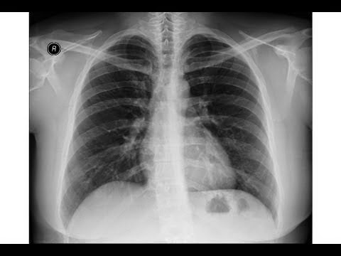
youtube viewer.Chest x-ray , pneumonia

Inguinal hernia

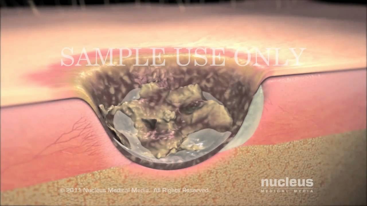
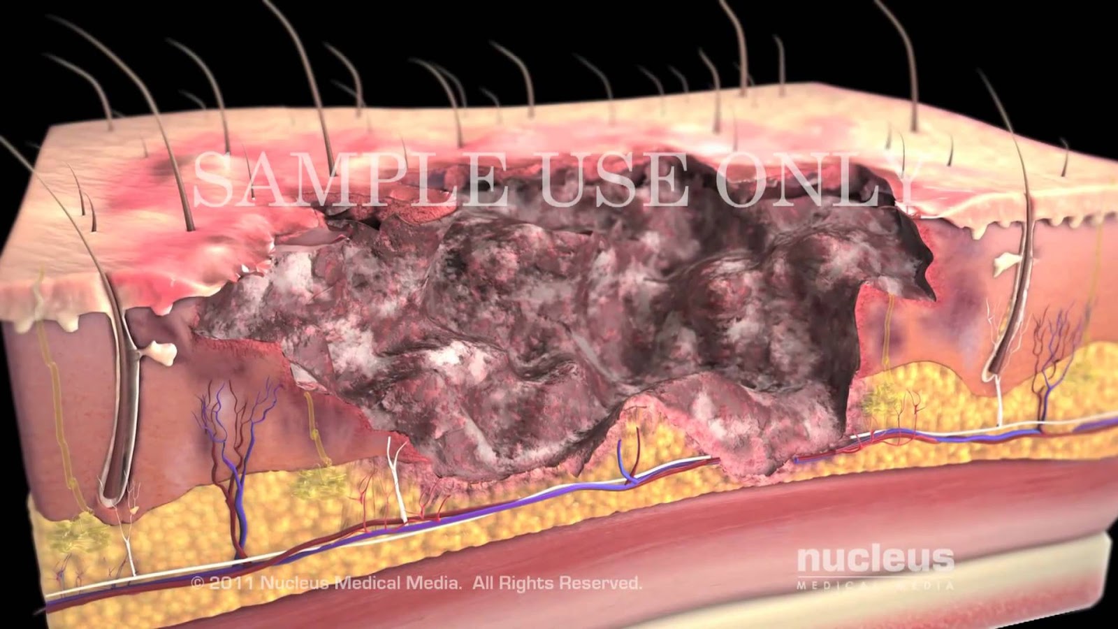
Surgical debridement
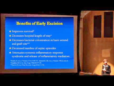
Burns: Classification and Treatment

A Medical Career - Why Not Pathology?

This five minute film interviews Pathologists about their chosen profession and highlights what a fascinating career Pathology can be.
Beyond the Data: Preventing Adverse Health Effects from Nanotechnology
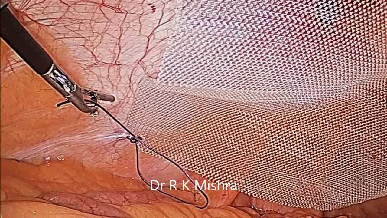
Laparoscopic Repair of Para Umbilical Hernia performed Dr R K Mishra
Subscribe to:
Posts (Atom)