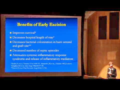
Burn Treatment - How to Achieve an LD50 of 98% TBSA Burn
Draining Bulla On Palm
 Here you will see drainage of bulla on right palm. You will see the
purulent material coming out when incision was given. Watch out whole
procedure of giving incision, cleaning wound, with normal saline,
hydrogen peroxide, betadiene, & removing dead skin.
Here you will see drainage of bulla on right palm. You will see the
purulent material coming out when incision was given. Watch out whole
procedure of giving incision, cleaning wound, with normal saline,
hydrogen peroxide, betadiene, & removing dead skin.Thoracoscopic (VATS) wedge resection of Fungal Ball (Mucormycosis) Left Upper Lobe
+wedge+resection+of+Fungal+Ball+(Mucormycosis)+Left+Upper+Lobe.jpg) This is an educational video demonstrating Thoracoscopic (VATS) wedge
resection of Fungal Ball (Mucormycosis) Left Upper Lobe by Prof. Arvind
Kumar and Dr. Belal Bin Asaf, Institute of Robotic Surgery at Sir Ganga
Ram Hospital, New Delhi, India.
This is an educational video demonstrating Thoracoscopic (VATS) wedge
resection of Fungal Ball (Mucormycosis) Left Upper Lobe by Prof. Arvind
Kumar and Dr. Belal Bin Asaf, Institute of Robotic Surgery at Sir Ganga
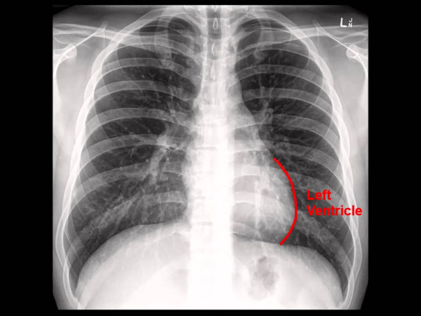
Ram Hospital, New Delhi, India. Chest X-ray: Analysis in a nutshell
Layers of the epidermis
 Lecture showing the 5 layers of the epidermis: basale, spinosum, granulosum, lucidum, corneum. The layers are showed on diagrams and in pictures taken by the use of light microscopy. The cells of the epidermis are also showed: Keratinocytes, Melanocytes, Merkell cells, Langherhans cells. Presents the difference between thick skin and thin skin.
Subscribe to:
Posts (Atom)