 Lecture showing the 5 layers of the epidermis: basale, spinosum, granulosum, lucidum, corneum. The layers are showed on diagrams and in pictures taken by the use of light microscopy. The cells of the epidermis are also showed: Keratinocytes, Melanocytes, Merkell cells, Langherhans cells. Presents the difference between thick skin and thin skin.
 Layers of the epidermis
Lecture showing the 5 layers of the epidermis: basale, spinosum, granulosum, lucidum, corneum. The layers are showed on diagrams and in pictures taken by the use of light microscopy. The cells of the epidermis are also showed: Keratinocytes, Melanocytes, Merkell cells, Langherhans cells. Presents the difference between thick skin and thin skin. Laparoscopic Lysis of Abdominal Adhesions

Basic Medical Pathology: Morphological Expressions of Cell Injury
 The video tape defines and describes injuries sustained by cells when
they are exposed to stress and are unable to adapt. The results of cell
injury are shown with photomicrographs, organ photographs, and electron
micrographs of normal and injured cells.
The video tape defines and describes injuries sustained by cells when
they are exposed to stress and are unable to adapt. The results of cell
injury are shown with photomicrographs, organ photographs, and electron
micrographs of normal and injured cells.Creating skin grafts for burn victims
 Biomedical engineers at Stevens are making more realistic skin grafts by adding hairs to artificial skin tissues.
Tuberculosis and Hypervascularity video - Animation by Cal Shipley, M.D.
 Infection of the lung by the tubercle bacillus with subsequent
inflammation and hypervascularity in the lungs. Formation of
hypervascular adhesions between the chest wall and lungs.
Infection of the lung by the tubercle bacillus with subsequent
inflammation and hypervascularity in the lungs. Formation of
hypervascular adhesions between the chest wall and lungs. Chest x-ray - Idiopathic Pulmonary Fibrosis
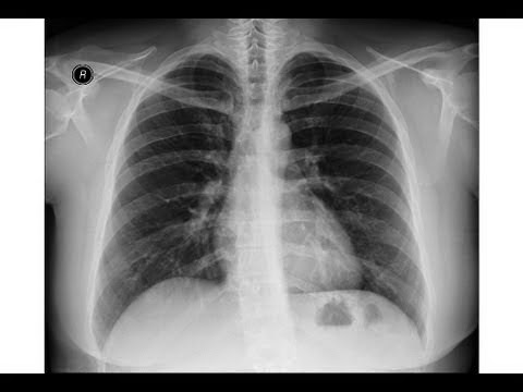 A brief video on IPF (IdiopathicPulmonary Fibrosis). X-ray with IPF is
courtesy of Dr Alex Maclennan and Normal Chest x-ray is courtesy of a
youtube viewer.
A brief video on IPF (IdiopathicPulmonary Fibrosis). X-ray with IPF is
courtesy of Dr Alex Maclennan and Normal Chest x-ray is courtesy of a
youtube viewer.Chest x-ray , pneumonia

Inguinal hernia

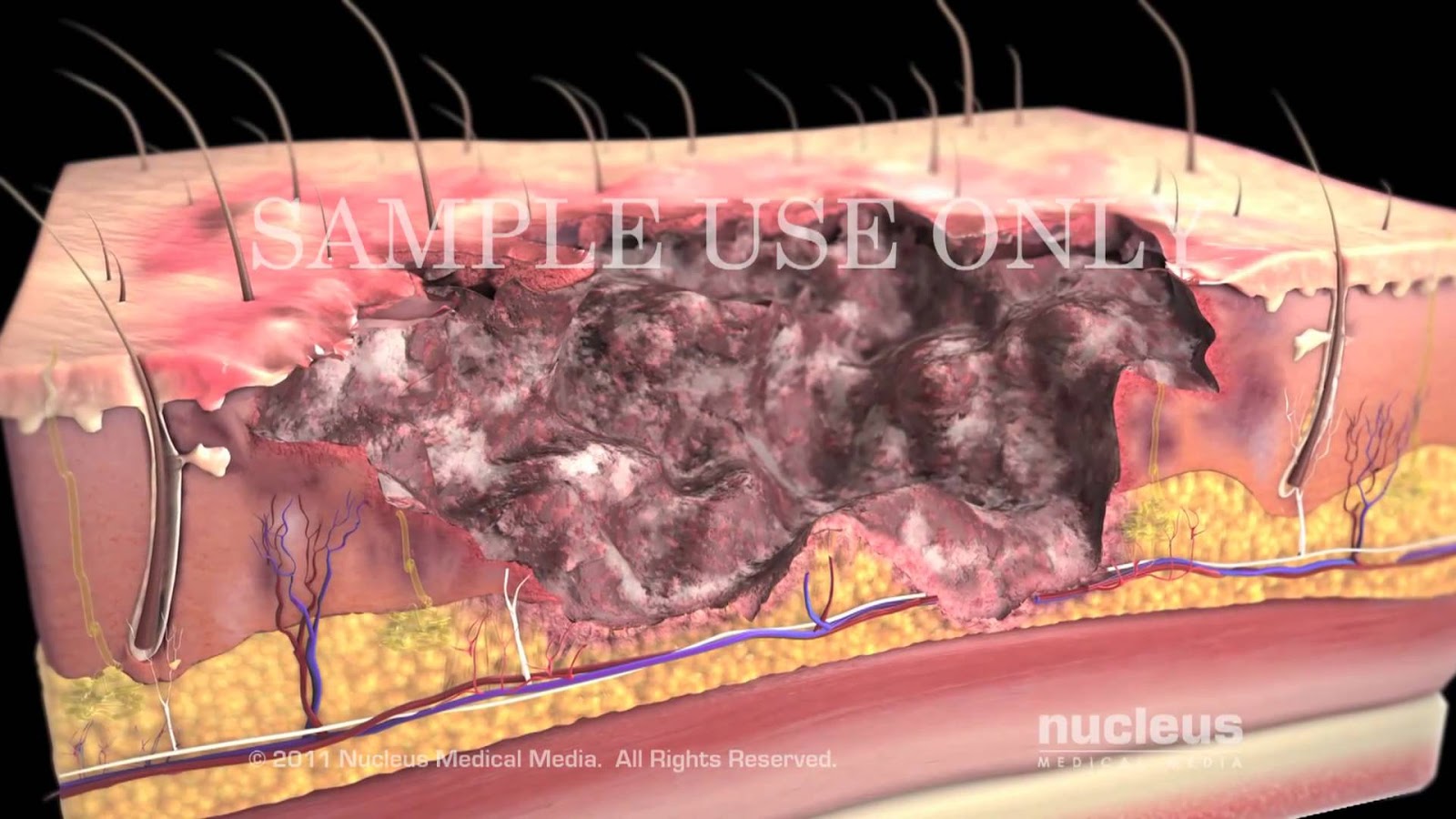
Surgical debridement
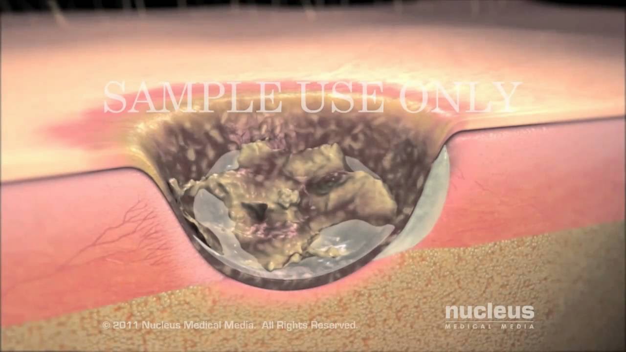
Burns: Classification and Treatment
Subscribe to:
Posts (Atom)